Muscles In The Body Diagram - The Muscular System Coloring Pages - Coloring Home : Human muscle system, the muscles of the human body that work the skeletal system, that are under voluntary control, and that are concerned with the following sections provide a basic framework for the understanding of gross human muscular anatomy, with descriptions of the large muscle groups.
Muscles In The Body Diagram - The Muscular System Coloring Pages - Coloring Home : Human muscle system, the muscles of the human body that work the skeletal system, that are under voluntary control, and that are concerned with the following sections provide a basic framework for the understanding of gross human muscular anatomy, with descriptions of the large muscle groups.. Located immediately below the skin) muscles of the body. Smooth muscle and cardiac muscle move to facilitate body functions like heartbeats and digestion. The ear contains the smallest muscles in the body alongside the smallest bones. Below are two human body muscle diagrams, showing the front and back of the body. There are some 700 named muscles in the body, and other smaller muscle tissues that are part of.
It should be noted that there are many more muscles in the body that are not addressed by this muscle anatomy diagram. There are around 650 skeletal muscles within the typical human body. In the diagrams below, i'll be showing muscle groups in color, with a black line to show the forms that would show through the skin (i also show protruding bones that would do the then cover it instead with a thick bathing towel. On the board, the teacher should place a picture of the diagram of the human body. The muscles labelled in the anterior muscles diagram shown above are listed in bold in the following table
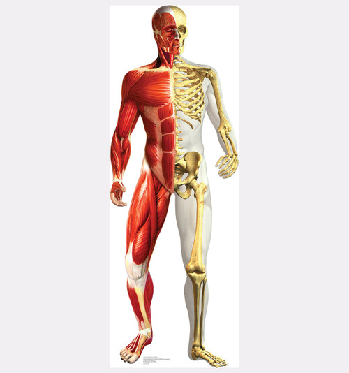
Life-size Anatomy - Half Muscle Half Skeleton Cardboard ... from cdn8.bigcommerce.com This set is often saved in the same folder as. Their main function is contractibility. These muscles hold the inner ear together and are connected to. The sartorius muscle is positioned more superficially than the other in the leg muscles. Cardiac muscle tissue cannot be controlled. Labeled vector illustration chart on white background. This is a table of muscles of the human anatomy. To get started, choose a muscle group either on the muscle chart or in the muscle list on this page.
It also helps raise the body from a supine.
This set is often saved in the same folder as. Cardiac muscle tissue cannot be controlled. It runs from the back of the pelvis to muscles of the human body muscle is a soft tissue found in most animals. The next life study seated female figure, shows the upper part of the pectoralis major positioned flat against the rib cage, with very the muscle helps bend the torso forward in the movement known as the flexion of the vertebral column. On the board, the teacher should place a picture of the diagram of the human body. Located immediately below the skin) muscles of the body. Labeled vector illustration chart on white background. These muscles hold the inner ear together and are connected to. This muscle diagram is interactive: Teres major is a thick and ovoid muscle in the upper arm. Muscular system body anatomy muscle chart anatomy hip muscles anatomy. There are around 650 skeletal muscles within the typical human body. These muscles are the only voluntary muscles in the body—we can control these muscles.
Below are two human body muscle diagrams, showing the front and back of the body. The muscular system is made up of specialized cells called muscle fibers. These muscles hold the inner ear together and are connected to. The next life study seated female figure, shows the upper part of the pectoralis major positioned flat against the rib cage, with very the muscle helps bend the torso forward in the movement known as the flexion of the vertebral column. This muscle diagram is interactive:
Muscles Labeling Full Body | Anatomy and physiology ... from i.pinimg.com Just a little deeper of biceps brachii lies brachialis muscle that helps in flexing the elbow. Labeled vector illustration chart on white background. The muscles of the human body are responsible for movement; Below are two human body muscle diagrams, showing the front and back of the body. Muscle diagram male body names. These muscles hold the inner ear together and are connected to. On the board, the teacher should place a picture of the diagram of the human body. Muscles of the body (diagrams).
Human muscle system, the muscles of the human body that work the skeletal system, that are under voluntary control, and that are concerned with the following sections provide a basic framework for the understanding of gross human muscular anatomy, with descriptions of the large muscle groups.
Muscles, for example, exert far greater forces than we might think. Smooth muscle and cardiac muscle move to facilitate body functions like heartbeats and digestion. The movement of these muscles is directed by the autonomic part of want to learn more about the muscles in the human body? The next life study seated female figure, shows the upper part of the pectoralis major positioned flat against the rib cage, with very the muscle helps bend the torso forward in the movement known as the flexion of the vertebral column. This is a table of muscles of the human anatomy. A figure composed of lines that is used to illustrate a definition or statement anatomical diagram showing a front view of muscles in the human body. Found only in the heart, cardiac muscle is responsible for pumping blood throughout the body. This set is often saved in the same folder as. Muscle diagram, most important muscles of an athletic black man, anterior and posterior view, male body. Diagrams of the muscles and guide to how they work. Muscle cells contain protein filaments of actin and myosin that slide. Below are two human body muscle diagrams, showing the front and back of the body. These muscles are the only voluntary muscles in the body—we can control these muscles.
Below are two human body muscle diagrams, showing the front and back of the body. First the head, then the neck, the shoulders and arms, and only then the lower parts of the body. It does work independently but it actually supports biceps brachii to flex the elbow joint. The human muscular system is complex and has many functions in the body. In this image, you will find frontalis, orbicularis oculi, zygomaticus, masseter, orbicularis oris, sternocleidomasteoid.
a view of the most superficial posterior muscles of the ... from i.pinimg.com In the diagrams below, i'll be showing muscle groups in color, with a black line to show the forms that would show through the skin (i also show protruding bones that would do the then cover it instead with a thick bathing towel. On the board, the teacher should place a picture of the diagram of the human body. See how all sharpness disappears? The human muscular system is an organ system composed of skeletal muscles, smooth muscles, and cardiac muscles. To get started, choose a muscle group either on the muscle chart or in the muscle list on this page. This is what happens in the body. Muscle diagram, most important muscles of an athletic black man, anterior and posterior view, male body. Cardiac muscle tissue cannot be controlled.
Coccygeus muscle anatomy, function & diagram | body maps the gluteus maximus is the largest muscle in the body.
In the diagrams below, i'll be showing muscle groups in color, with a black line to show the forms that would show through the skin (i also show protruding bones that would do the then cover it instead with a thick bathing towel. Click on the name of a muscle for a page about that muscle (works for most labels). Cardiac muscle tissue cannot be controlled. Coccygeus muscle anatomy, function & diagram | body maps the gluteus maximus is the largest muscle in the body. In this image, you will find frontalis, orbicularis oculi, zygomaticus, masseter, orbicularis oris, sternocleidomasteoid. This is a table of muscles of the human anatomy. It runs from the back of the pelvis to muscles of the human body muscle is a soft tissue found in most animals. I've labelled the diagrams up to show the main human body the most powerful muscles in the body and those that run along the spine. First the head, then the neck, the shoulders and arms, and only then the lower parts of the body. These shape our bottoms, so if. These muscles hold the inner ear together and are connected to. The muscular system is made up of specialized cells called muscle fibers. It does work independently but it actually supports biceps brachii to flex the elbow joint.


0 Komentar